
Application of PET/CT using 18F-FDG in myocardial infarction, lymphoma and colorectal cancer patients
Keywords:
colorectal cancer, consecutive lymphoma patients, myocardial infarction patients, PET/CT, 18F-FDGAbstract
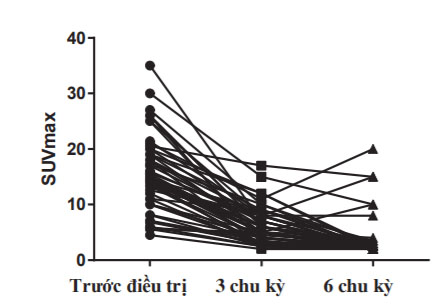
This paper aimed (1) to demonstrate the benefit of 18F-FDG PET/CT in myocardial viability assessement for post-myocardial infarction patients; (2) to evaluate the role of 18F-FDG PET/CT in staging and distinguishing between responders and non-responders of lymphoma patients to standard treatment; and (3) to determine the value of 18F-FDG PET/CT in staging of patients with preoperative and postsurgical colorectal cancer. Materials and methods: myocardial viability assessment was underwent in 69 post-myocardial infarction patients with left ventricular dysfunction by SPECT myocardial perfusion and 18F-FDG PET/CT scan to assess myocardial viability. Left ventricular functions by echocardiography in 30 patients with myocardial hibernating were monitored at 6 and 12 months after coronary revascularization. A total of 60 consecutive lymphoma patients were newly diagnosed, and all these patients were undergone 18F-FDG PET/CT scan before chemotherapy, after 3-cycle and 6-cycle treatment. A total of 70 patients who was diagnosed colorectal tumor by endoscopy with histology. The patients were scanned with 18F-FDG PET⁄CT and conventional diagnostic imaging. The reference method selected was pathological histology when possible. 40 patients suspicious for colorectal cancer recurrences based on clinical symptoms or serum carcinoembryonic antigen (CEA) level were recruited to the study. All patients underwent conventional imaging and 18F-FDG PET/CT. Sensitivity (SEN), specificity (SPE), positive predictive value (PPV) and negative predictive value (NPV) were calculated using either histopathology or follow-up imaging as the standard of reference. The results showed that, myocardial hibernating was detected on 18F-FDG PET imaging in 29% of 69 patients with postmyocardial infarction. EF% was improved significantly in groups of patients with large hibernating defect who was revascularized from 40.5±5.18% to 48.3±4.05 after 12 months. 18F-FDG PET/CT could detect more nodal and extranodal lymphomas compared to CT imaging. After 3-cycle chemotherapy, the results showed that 53.5% patients with complete response (CR), 33.3% with partial response who was continuing the previous regime of chemotherapy (PR). Meanwhile, 11.8% who had PET-positive received an alternative chemotherapy regime. There were significant relationships between 18F-FDG PET/CT imaging after 3-cycle and 6-cycle chemotherapy in lymphoma patients. Regarding to detection of regional lymph node metastases in pre-surgical colorectal cancer patients, 18F-FDG PET/CT showed higher sensitivity (78%) and specificity (84%) than CT alone (61% and 69%, respectively). For distant metastase assessment, the sensitivity and specificity of 18F-FDG PET/CT were 100% in all patients who had liver lesions. Regarding to detection of local recurrences, 18F-FDG PET/CT showed sensitivity of 100%; specificity 67%; PPV 88%; NPV 100%. The SEN, SPE, PPV, NPV of conventional imaging was 43%, 100%, 100%, 43%, respectively. The value of CEA level was 64%, 67%, 44%, 81% in SEN, SPE, PPV and NPV respectively. The SEN, SPE, PPV, NPV of 18F-FDG PET/CT in detecting lymph node metastases were 100%, 88%, 80% and 100% which are higher than those values of CT. The results suggest that outcome may be benefited using 18F-FDG PET in an experienced center with ready access to 18F-FDG and integration with imaging and revascularization teams. 18F-FDG PET/CT has high potential to help clinicians make decision for further treatment of lymphoma patients. 18F-FDG PET/CT is superior to conventional imaging in pre-surgical colorectal cancer, and 18F-FDG PET/CT has high accuracy in the assessment of local recurrences and metastases impacting significantly in colorectal cancer patient management.
Classification number
3.2
Downloads
Published
Received: 18 January 2016; accepted: 26 February 2016



